Posterior Rib Cage Muscles - What Is The Role Of Intercostal Muscles In Respiration And Where Are They Found Quora. It is made up of 12 pairs of ribs. Rib 2 is thinner and longer than rib 1, and has two articular facets on the head as normal. Read more below to learn what may be causing your rib pain and when to seek treatment. Look for clues from landmarks and muscle attachments that will tell you exactly where the rib cage is. There are also some smaller, deeper muscles which lie against the rib cage.
These muscles attach the upper limb to the axial skeleton of the trunk and support the thoracic cage. The brachial chain muscle on the left is opposed by the left posterior back muscles (pec), lower trap, serratus anterior, external rib rotators and right internal abdominal obliques. The accessory muscles include the scalenes and the sternocleidomastoid muscles in the neck, the serratus anterior, and the pectoral muscles in the upper trunk, the upper trapezius and latissimus dorsi muscles of the trunk, and the erector spinae muscles of the back. It has a roughened area on its upper surface, from which the serratus anterior muscle originates. In addition to aiding in breathing, the intercostal muscles also help stabilize the rib cage as the upper body twists or bends forward, backward, or to the side.
Muscles In The Thoracic Region That Help You Breathe Dummies from www.dummies.com Rib cage pain can be caused. Intercostal muscles are muscles that present within the rib cage. Our ribcage exists to protect the heart and lungs. The brachial chain muscle on the left is opposed by the left posterior back muscles (pec), lower trap, serratus anterior, external rib rotators and right internal abdominal obliques. There are also some smaller, deeper muscles which lie against the rib cage. Diaphragm, iliacus, psoas, tfl, vastus lateralis, biceps femoris Some evidence supports 11 a relationship between trunk muscle activity and posterior chain muscle movement. Learn vocabulary, terms, and more with flashcards, games, and other study tools.
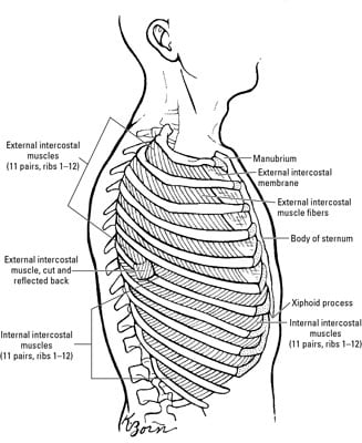
The fibres pass superolaterally to insert into the internal surface of costal cartilages of ribs two to six. The middle and upper part of your spine is called the thoracic region and it helps to support your upper body. Construct a robo skelly rib cage and the pelvis using the bucket method. The accessory muscles include the scalenes and the sternocleidomastoid muscles in the neck, the serratus anterior, and the pectoral muscles in the upper trunk, the upper trapezius and latissimus dorsi muscles of the trunk, and the erector spinae muscles of the back. The transversus thoracic muscles originate from the posterior surface of the xiphoid process and the lower part of the body of the sternum. Related posts of rib cage diagram with organs abdominal cavity chart. Rib cage pain may be sharp, dull, or achy and felt at or below the chest or above the navel on either side. It is made up of 12 pairs of ribs. Some evidence supports 11 a relationship between trunk muscle activity and posterior chain muscle movement. Learn vocabulary, terms, and more with flashcards, games, and other study tools. External intercostals muscle are the outermost layer lies directly under the skin originate from the lower border of rib above run obliquely and insert into the upper border of the rib below. An inhalation is accomplished when the muscular diaphragm, at the floor of the thoracic cavity, contracts and flattens, while the contraction of intercostal muscles lift the rib cage up and out. According to the book clinical anatomy of the spine, intercostal muscles also influence the spine.
Try to be as accurate as you can with them. This condition is characterized by inflammation of the cartilage in the rib cage. Start studying tx/rib cage anatomy. Rib cage, in vertebrate anatomy, basketlike skeletal structure that forms the chest, or thorax, and is made up of the ribs and their corresponding attachments to the sternum (breastbone) and the vertebral column.the rib cage surrounds the lungs and the heart, serving as an important means of bony protection for these vital organs.in total, the rib cage consists of the 12 thoracic vertebrae and. The transversus thoracic muscles originate from the posterior surface of the xiphoid process and the lower part of the body of the sternum.
Muscles Of Expiration Thoracic And Abdominal Chapter 2 Flashcards Quizlet from o.quizlet.com Look for clues from landmarks and muscle attachments that will tell you exactly where the rib cage is. In addition to aiding in breathing, the intercostal muscles also help stabilize the rib cage as the upper body twists or bends forward, backward, or to the side. It has a roughened area on its upper surface, from which the serratus anterior muscle originates. The intercostals (external, internal and innermost), subcostals, and transversus thoracis. There are some other muscles that do not comprise the thoracic wall, but do attach to it. Costochondritis or tietze's syndrome is another common cause of rib cage pain. The thoracic cage (rib cage) is the skeletal framework of the thoracic wall, which encloses the thoracic cavity. Oftentimes, pain under the rib cage is not serious and may be associated with minor conditions like indigestion, gas troubles, or strained muscles.
Costochondritis or tietze's syndrome is another common cause of rib cage pain.
The rib cage is a bony structure found in the chest (thoracic cavity). Costochondritis or tietze's syndrome is another common cause of rib cage pain. Each are symmetrically paired on a right and left side. It has a roughened area on its upper surface, from which the serratus anterior muscle originates. Rib cage pain may be sharp, dull, or achy and felt at or below the chest or above the navel on either side. Our ribcage exists to protect the heart and lungs. These muscles act to change the volume of the thoracic cavity during respiration. These muscles attach the upper limb to the axial skeleton of the trunk and support the thoracic cage. The human rib cage is a component of the human respiratory system. It may occur after an obvious injury or without explanation. Don't just draw a generic rib cage shape in there. The brachial chain muscle on the left is opposed by the left posterior back muscles (pec), lower trap, serratus anterior, external rib rotators and right internal abdominal obliques. Anterior interior chain (aic) muscles:
It encloses the thoracic cavity, which contains the lungs. Muscle anatomy amazon 12 photos of the muscle anatomy amazon amazon muscle anatomy poster, muscle anatomy amazon, muscle anatomy model amazon, muscle trigger point anatomy amazon, human muscles, amazon muscle anatomy poster, muscle anatomy amazon, muscle anatomy model amazon, muscle trigger point anatomy amazon Costochondritis or tietze's syndrome is another common cause of rib cage pain. Techniques aimed at the diaphragm have been used to increase movement in the rib cage and the spine 9 , 10. The brachial chain muscle on the left is opposed by the left posterior back muscles (pec), lower trap, serratus anterior, external rib rotators and right internal abdominal obliques.
Getting To The Bottom Of Rib Cage Pain Nydnrehab Com from nydnrehab.com The transversus thoracic muscles originate from the posterior surface of the xiphoid process and the lower part of the body of the sternum. It may occur after an obvious injury or without explanation. Consist of three layers of muscles external, internal, and innermost layer they combine to fill the space between the ribs. External intercostals muscle are the outermost layer lies directly under the skin originate from the lower border of rib above run obliquely and insert into the upper border of the rib below. The human rib cage is a component of the human respiratory system. Diaphragm, iliacus, psoas, tfl, vastus lateralis, biceps femoris Post your work in the anatomy for artists. Some evidence supports 11 a relationship between trunk muscle activity and posterior chain muscle movement.
Related posts of rib cage diagram with organs abdominal cavity chart.
Rib pain or pain in the chest wall that feels like it comes from a rib may be caused by traumatic injury, muscle strain, joint inflammation, or chronic pain, and ranges in severity. These muscles attach the upper limb to the axial skeleton of the trunk and support the thoracic cage. Abdominal cavity chart 14 photos of the abdominal cavity chart abdominal cavity cancer, abdominal cavity contains, abdominal cavity diagram picture, abdominal cavity pain, abdominal cavity quadrants, abdominal cavity regions, air in abdominal cavity, fluid buildup in abdominal cavity, stomach, abdominal cavity cancer. There are also some smaller, deeper muscles which lie against the rib cage. Between each rib lie several layers of intercostal muscles that are responsible for expanding and shrinking the rib cage when we breathe. Muscle anatomy amazon 12 photos of the muscle anatomy amazon amazon muscle anatomy poster, muscle anatomy amazon, muscle anatomy model amazon, muscle trigger point anatomy amazon, human muscles, amazon muscle anatomy poster, muscle anatomy amazon, muscle anatomy model amazon, muscle trigger point anatomy amazon It may occur after an obvious injury or without explanation. The fibres pass superolaterally to insert into the internal surface of costal cartilages of ribs two to six. Don't just draw a generic rib cage shape in there. Diaphragm, iliacus, psoas, tfl, vastus lateralis, biceps femoris Intercostal muscles are muscles that present within the rib cage. Rib cage pain can be caused. The human rib cage is a component of the human respiratory system.
Rib cage pain can be caused rib cage muscles. The human rib cage is made up of 12 paired rib bones;
Share :
Post a Comment
for "Posterior Rib Cage Muscles - What Is The Role Of Intercostal Muscles In Respiration And Where Are They Found Quora"


Post a Comment for "Posterior Rib Cage Muscles - What Is The Role Of Intercostal Muscles In Respiration And Where Are They Found Quora"